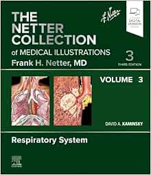
The Netter Collection of Medical Illustrations: Respiratory System, Volume 3, 3rd Edition (True Publisher)
$37.00
Breathe clarity into your medical studies with The Netter Collection of Medical Illustrations: Respiratory System, Volume 3 (3rd Edition, 2024). This updated edition combines Frank H. Netter’s legendary medical artwork with concise, clinically relevant insights, making it an essential reference for students, pulmonologists, thoracic surgeons, and anyone involved in the diagnosis and treatment of respiratory diseases.
Description
A Definitive Visual Guide to the Human Respiratory System
As part of Elsevier’s prestigious Netter Green Book Collection, Respiratory System, Volume 3 (3rd Edition, 2024) delivers a comprehensive visual and clinical exploration of the lungs, airways, pleura, and associated structures. Featuring the meticulous illustrations of Frank H. Netter, MD, this edition is enhanced with modern updates on diagnostics, disease mechanisms, and clinical procedures—bridging classic anatomical education with today’s respiratory medicine.
Whether you’re a student grasping the fundamentals of pulmonary physiology or a seasoned practitioner needing a trusted visual reference, this book offers a high-yield, visually immersive learning experience.
Target Audience:
- Medical and respiratory therapy students
- Pulmonology and thoracic surgery residents
- Clinicians in internal medicine and critical care
- Nurse practitioners, PAs, and respiratory therapists
- Medical educators and anatomy instructors
Key Features & Highlights:
- Legendary Netter Illustrations: Accurate, full-color drawings that capture the intricacies of respiratory anatomy and disease.
- Clinically Integrated Content: Visuals are paired with updated text covering pathophysiology, imaging, diagnostics, and treatment strategies.
- Up-to-Date Medical Insights: Reflects the latest in respiratory care, including COVID-19 impacts, interstitial lung diseases, and thoracic procedures.
- Comprehensive System Coverage: Includes core chapters on nasal passages, trachea, lungs, bronchial tree, pleural cavities, and diaphragm.
- Educational Versatility: Ideal for academic study, exam preparation, patient communication, and surgical planning.
Topics Covered:
Includes core chapters on:
- Upper and lower airway anatomy
- Pulmonary vasculature and gas exchange
- Pleural membranes and thoracic cavity structures
- Common respiratory diseases such as asthma, COPD, pneumonia, and lung cancer
- Surgical and diagnostic approaches including bronchoscopy and thoracentesis
About the Author:
Under Elsevier’s expert editorial leadership, this volume builds on the foundational work of Dr. Frank H. Netter, whose illustrations have educated generations of healthcare professionals with unparalleled anatomical precision and clarity.
Technical Details:
- File Format: PDF
- File Size: Approx. 135 MB
- Language: English
- Device Compatibility: Fully compatible with iOS, Android, Windows, and macOS devices—optimized for tablets, laptops, and mobile reading
Frequently Asked Questions:
Q1: Does this volume include clinical pathologies and disease-specific illustrations?
Yes. This edition combines detailed anatomical drawings with depictions of major respiratory diseases and related clinical interventions, making it useful for both learning and practice.
Q2: Is this ebook suitable for USMLE or board exam preparation?
Absolutely. The book’s comprehensive visuals and concise explanations make it an excellent supplemental resource for medical exams, especially those involving anatomy and respiratory physiology.
Additional information
| Publisher |
Elsevier |
|---|---|
| Published Year |
2024 |
| Language |
English |
| ISBN |
9780323881272 |
| File Size |
54.1 MB |
| Edition |
3 |


Reviews
There are no reviews yet.